Active AFM Cantilevers And The New Research They Enable
- 19 May 2026
- Volume 30
- NANOscientific Magazine, 2026
Prof. Georg E. Fantner, École Polytechnique Fédérale de Lausanne (EPFL), Lausanne, Switzerland
Introduction: Beyond Miniaturization
Since its introduction in 1986, the atomic force microscope (AFM) has evolved largely through advances in cantilever design and microfabrication. Early progress was driven by miniaturization, making cantilevers smaller, faster, and more precise. This strategy dramatically increased imaging speed and force sensitivity, enabling high-speed AFM and expanding the technique into liquid environments and dynamic biological systems.
However, miniaturization alone eventually encounters practical and physical limits. Extremely small cantilevers are difficult to handle and operate reliably, particularly in applications requiring larger scan ranges or stable force control. Further increases in resonance frequency yield diminishing returns when bandwidth becomes limited by the quality factor (Q) and intrinsic material losses.
In his recent presentation, Professor Georg Fantner revisits cantilever design from a different perspective: rather than focusing exclusively on size reduction, he explores how material choice, intrinsic damping, and multilayer microfabrication can fundamentally alter cantilever dynamics and enable new and enhanced measurement modalities. His work demonstrates that innovation in AFM hardware, particularly in cantilever engineering, continues to unlock new scientific capabilities, from high-speed air imaging to self-sensing detection, in-SEM correlative metrology, and even approaches toward three-dimensional tomography.
Cantilever Dynamics: Rethinking Speed and Bandwidth
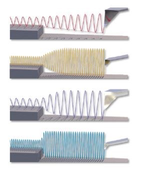
In dynamic (tapping-mode) AFM, the response time of the cantilever determines imaging speed and force control. When the cantilever encounters a change in tip-sample interaction, it must transition to a new steady-state oscillation. The time required for this transition sets the imaging bandwidth.
Traditionally, bandwidth improvement has been pursued by increasing resonance frequency through miniaturization. Since the response time scales approximately with the ratio of Q factor to resonance frequency, reducing cantilever size increases frequency and therefore shortens response time.
Fantner highlights a complementary strategy: instead of only increasing resonance frequency, one can also reduce the intrinsic Q factor. Because the bandwidth scales with f₀/Q, modifying material properties to intrinsically lower Q can achieve comparable, or even superior, dynamic response without extreme miniaturization.
This shift in perspective leads directly to reconsidering the materials used in cantilever fabrication.
Polymer Cantilevers: Intrinsically Faster Dynamics
Most commercial cantilevers are fabricated from silicon or silicon nitride, materials that provide high mechanical stability and low intrinsic mechanical losses. However, these same properties contribute to relatively high Q factors, particularly in air and vacuum.
Fantner’s group investigated polymer-based cantilevers, specifically using the photodefinable epoxy SU-8. While polymers typically exhibit greater intrinsic mechanical losses, this characteristic becomes advantageous in dynamic AFM because it lowers Q and improves transient response.1,2
By fabricating cantilevers with identical geometries in both conventional materials and SU-8, the team demonstrated:
- Reduced Q factors in polymer cantilevers.
- Significantly improved dynamic response in air.
Small SU-8 cantilevers achieved imaging rates on the order of one image per second in air for typical scan conditions, a performance typically associated with high-speed AFM in liquid. Moreover, these improvements were achieved without sacrificing force control stability.
An important secondary benefit is gentler imaging. Faster dynamic response reduces feedback lag, enabling lower interaction forces and improved topographical fidelity. High-resolution images (e.g., 26 Megapixels acquired within practical acquisition times) demonstrate that speed enhancements translate directly into better force regulation and data quality.
Rather than simply making cantilevers smaller, Fantner’s work shows that selecting materials with tailored intrinsic loss properties can fundamentally reshape AFM performance.
Self-Sensing Cantilevers: Overcoming Optical Lever Limitations
Despite decades of innovation, the optical beam deflection method
remains the dominant AFM detection scheme. While highly sensitive,
optical detection introduces constraints:
- Alignment complexity.
- Space requirements.
- Incompatibility with confined geometries.
- Difficulty operating in opaque liquids.
- Limited integration with other instruments.
Self-sensing cantilevers, particularly piezoresistive designs, offer a compact
alternative. However, they historically suffer from lower sensitivity.
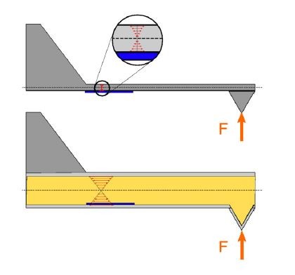
The reason lies in what is being measured. Optical detection measures
deflection or angular change, whereas piezoresistive detection measures
strain. Since strain is related to curvature (the second derivative
of displacement), thinning the cantilever, which lowers the spring
constant and improves optical sensitivity, reduces strain sensitivity because
the distance between the sensing elements and the neutral axis
decreases.
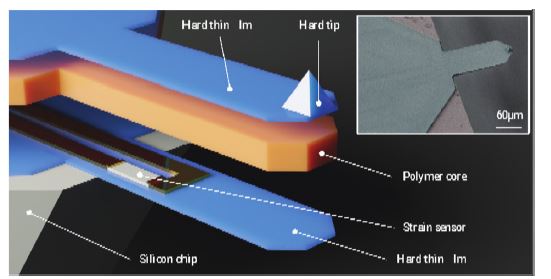
Fantner’s solution is a trilayer cantilever architecture3:
- Thin silicon nitride layers with embedded piezoresistive sensing elements.
- Thick polymer core forming the structural bulk.
By embedding sensing elements farther from the neutral axis while
maintaining low spring constants through polymer compliance, the design
significantly increases strain sensitivity4.
Two-Wafer Microfabrication
Fabrication involves a two-wafer bonding process:
- One wafer defines silicon or silicon nitride tips.
- The second wafer carries the piezoresistive sensing elements and interconnects.
- After high-temperature processing, a polymer core is introduced.
- The wafers are then bonded, followed by micromachining steps to define and release trilayer cantilevers.
All sensing traces are encapsulated within the cantilever, making the device robust in liquid environments and chemically harsh conditions.
The result is a self-sensing cantilever with force noise approaching that of optical detection and operation close to the thermal noise limit.
Actuation Integration: Toward Faster Off-Resonance Imaging
The flexibility of microfabrication enables the integration of both sensing and actuation. Fantner’s cantilevers incorporate integrated actuation (e.g., electrothermal and piezoelectric), allowing direct excitation of cantilever motion5,6.
Direct actuation reduces spurious resonances associated with external piezo excitation and can improve response speed by up to two orders of magnitude.
This capability enables rapid off-resonance imaging. In this mode, the cantilever is driven below resonance, and the tip-sample interaction is directly probed. With integrated actuation, mechanical property mapping can be performed on the order of one to two seconds per image while preserving quantitative force information.
Correlative AFM Inside the SEM
The compactness of self-sensing cantilevers enables integration into confined environments. Fantner’s group developed an AFM system capable of operating inside a scanning electron microscope (SEM)7, later commercialized in collaboration with Quantum Design.
Modern SEM chambers provide limited space above the sample, making optical detection impractical. Self-sensing cantilevers eliminate the need for laser alignment and bulky optical components.
This integration enables:
- Direct navigation to regions of interest.
- Correlative topography and mechanical mapping.
- Imaging of irregular or structured samples.
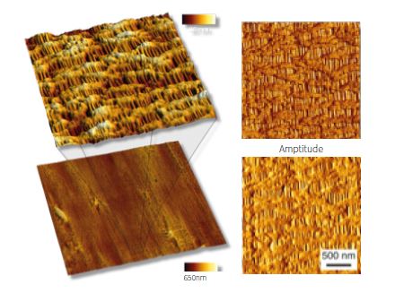
For example, the mechanical characterization of self-healing composite polymers was performed within the SEM. AFM provided nanoscale stiffness contrast across structural fibers and thermoplastic regions, information not directly accessible through SEM imaging alone.
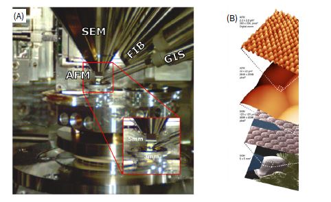
Three-Dimensional AFM: Toward Tomography
AFM is inherently a surface technique. Extending it into the third dimension has long been a challenge. Fantner’s solution combines AFM with focused ion beam (FIB) milling inside a dual-beam FIB-SEM instrument. Sequential slicing exposes successive surfaces, which are then imaged mechanically.
In many cases, topography changes little between slices, while stiffness mapping reveals internal heterogeneity. In rubber samples, carbon filler particles appear as stiff inclusions within a soft matrix, enabling three-dimensional stiffness mapping.
This approach extends AFM from a surface profiler toward a volumetric mechanical characterization tool.
Impact and Outlook
Fantner’s work underscores a central theme: microfabrication remains a key enabling technology for AFM evolution. By rethinking materials, architecture, and integration strategies, cantilevers become multifunctional platforms rather than passive mechanical probes.
Key contributions include:
- Polymer cantilevers with intrinsically lower Q for faster air imaging.
- Trilayer self-sensing designs with improved strain sensitivity.
- Integrated actuation for rapid off-resonance imaging.
- Integration of AFM within SEM and FIB systems.
- Three-dimensional stiffness tomography.
Nearly four decades after the introduction of AFM, the cantilever remains central to innovation. Rather than being limited by physical constraints, creative microfabrication strategies continue to expand the scope of what AFM can measure, mechanically, electrically, and volumetrically.
As AFM increasingly intersects with electron microscopy, cryogenic techniques, and multimodal characterization, the cantilever evolves from a simple mechanical beam into a multifunctional nanoscale sensor.
The field’s future may lie not in making cantilevers smaller, but in making them smarter.

Georg E. Fantner received his MS from the Graz University of Technology (2003) and his PhD from the University of California, Santa Barbara (2006). Following a postdoctoral position in the Biomolecular Materials Lab at the Massachusetts Institute of Technology, he joined the École Polytechnique Fédérale de Lausanne in 2010. He leads the Laboratory for Bio- and Nano-Instrumentation and serves as co-director of the Institute of Bioengineering.
His research focuses on developing advanced technologies for measuring and manipulating nanoscale structures, particularly atomic force microscopy instrumentation, with applications spanning materials science, nanotechnology, and the life sciences. His work has been published in leading journals including Nature, Nature Materials, Nature Physics, Nature Nanotechnology, Nature Cell Biology, Nature Microbiology, Nature Communications, Nano Letters, and Science, and has been featured in popular science publications.
Prof. Fantner is co–Editor-in-Chief of Microsystem Technologies (Springer Nature) and serves as scanning probe microscopy editor for Microscopy and Microanalysis. He holds several patents in nanotechnology and is co-founder of two nanotechnology companies. He is also active in open hardware initiatives to promote academic knowledge sharing and serves as president of the EPFL Open Science Strategic Committee and the ETH Domain Open Research Data Steering Committee.
References
- Adams, J. D. et al. Harnessing the damping properties of materials for high-speed atomic force microscopy. Nat. Nanotechnol. 11, 147–151 (2016).
- Hosseini, N. et al. Integration of sharp silicon nitride tips into high-speed SU8 cantilevers in a batch fabrication process. Beilstein J. Nanotechnol. 10, 2357–2363 (2019).
- Hosseini, N. et al. Batch Fabrication of Multilayer Polymer Cantilevers with Integrated Hard Tips for High-Speed Atomic Force Microscopy. in 2019 20th International Conference on Solid-State Sensors, Actuators and Microsystems and Eurosensors XXXIII, TRANSDUCERS 2019 and EUROSENSORS XXXIII (2019). doi:10.1109/TRANSDUCERS.2019.8808606.
- Hosseini, N. et al. A polymer–semiconductor–ceramic cantilever for high-sensitivity fluid-compatible microelectromechanical systems. Nat. Electron. 7, 567–575 (2024).
- Neuenschwander, M., Andany, S. H., Kangul, M., Hosseini, N. & Fantner, G. E. Self-Actuated Polymer-Based Cantilevers with Sharp Silicon Tips for High-Speed Atomic Force Microscopy. in 2021 21st International Conference on Solid-State Sensors, Actuators and Microsystems (Transducers) 22–25 (IEEE, 2021). doi:10.1109/Transducers50396.2021.9495715.
- Neuenschwander, M. et al. Fabrication of fluid-compatible trilayer cantilevers with integrated piezoelectric actuators. Sens. Actuators Phys. 383, 116217 (2025).
- Andany, S. H. et al. An atomic force microscope integrated with a helium ion microscope for correlative nanoscale characterization. Beilstein J. Nanotechnol. 11, 1272–1279 (2020).
Category